Understanding Physical Therapy Protocols in Post-Surgical Recovery

Understanding Physical Therapy Protocols in Post-Surgical Recovery

Explore the essential physical therapy protocols in post-surgical recovery to enhance healing and restore mobility. This guide provides valuable insights into effective rehabilitation strategies tailored for various surgical procedures.

How does the application of neuromuscular electrical stimulation (NMES) fit into post-surgical rehabilitation protocols for knee surgeries?

Neuromuscular electrical stimulation (NMES) plays a significant role in post-surgical rehabilitation protocols for knee surgeries, particularly after procedures like anterior cruciate ligament (ACL) reconstruction or total knee arthroplasty. By delivering controlled electrical impulses to the muscles around the knee joint, NMES promotes muscle activation and contraction, which helps counteract postoperative muscle atrophy and weakness commonly seen after surgery. This technique enhances neuromuscular re-education by stimulating both slow-twitch and fast-twitch fibers, facilitating improved strength gains during early recovery phases when patients might struggle with voluntary contractions due to pain or swelling. Additionally, NMES aids in reducing joint stiffness by improving circulation through increased blood flow to the affected area while also promoting quicker healing of soft tissues surrounding the surgical site. The integration of NMES into a comprehensive rehabilitation plan often includes parameters such as frequency, duration, and intensity tailored specifically for each patient's needs; this customization ensures optimal therapeutic effects without causing further discomfort or aggravation of injuries. Furthermore, using NMES alongside traditional physical therapy techniques fosters better outcomes regarding range of motion restoration and functional mobility in daily activities post-surgery. In summary, incorporating neuromuscular electrical stimulation into post-operative care not only supports effective recovery trajectories but also empowers patients on their journey towards regaining full functionality following knee surgeries.

Orthopedic physical therapy can significantly enhance recovery from musculoskeletal injuries by improving strength and flexibility. By engaging in these specialized programs, individuals often experience reduced pain and a quicker return to daily activities. To explore how orthopedic physical therapy can benefit you, visit notes about physical therapy and how it is used for post-surgery recovery

What role do joint mobilization techniques play in addressing range of motion deficits following rotator cuff repair?

Joint mobilization techniques play a significant role in enhancing range of motion deficits that often occur after rotator cuff repair surgery. These specialized manual therapy methods involve the application of controlled movements to the joints and surrounding tissues, which can help reduce stiffness, improve flexibility, and restore normal function in the shoulder area. By focusing on both passive and active movement strategies, joint mobilization enables patients to gradually regain their mobility while minimizing pain through gentle stretching of tight muscles and fascia around the shoulder joint. This process is essential for breaking down scar tissue that may form post-surgery, ultimately aiding in better alignment of bones within the glenohumeral joint. Additionally, these techniques work synergistically with rehabilitation exercises designed for strength building by preparing soft tissues for more intensive training without causing further injury or strain during recovery phases. As such, incorporating joint mobilization into post-operative care plans not only enhances overall patient outcomes but also fosters greater independence as individuals return to daily activities following rotator cuff repairs.

In what ways can proprioceptive training be integrated into physical therapy plans after anterior cruciate ligament (ACL) reconstruction?

Proprioceptive training can be integrated into physical therapy plans after anterior cruciate ligament (ACL) reconstruction through various methods that enhance balance, coordination, and joint awareness. One effective approach involves incorporating specific exercises like single-leg stands on unstable surfaces such as balance boards or foam pads to challenge the proprioceptors in the ankle and knee joints, thereby improving stability and preventing re-injury. Additionally, activities such as agility drills using cones or mini-hurdles can be introduced gradually to develop neuromuscular control while simulating sports-specific movements. Utilizing tools like resistance bands during functional tasks helps strengthen surrounding musculature while simultaneously enhancing proprioceptive feedback from these muscles. Furthermore, implementing closed kinetic chain exercises—like squats or leg presses—promotes weight-bearing stabilization essential for restoring function post-surgery; this method not only engages multiple muscle groups but also reinforces proper movement patterns critical for future athletic performance. Incorporating visual cues during rehabilitation sessions encourages cognitive engagement with motor learning principles that further improve dynamic balance skills necessary for safe return-to-play protocols following ACL injury recovery. Moreover, integrating plyometric training progressively allows patients to develop explosive strength combined with reactive abilities required in sport contexts while ensuring they maintain adequate proprioception throughout their rehabilitation journey to optimize outcomes effectively without compromising safety measures involved in their rehab process.

How is graded exposure utilized to manage pain and anxiety during early-phase recovery from total hip arthroplasty?

Graded exposure is a therapeutic technique that plays a crucial role in managing pain and anxiety during the early-phase recovery from total hip arthroplasty, which involves replacing damaged or worn-out parts of the hip joint. This method focuses on gradually increasing activities and movements that patients may find challenging due to their fear of pain or re-injury. By starting with simple, less intimidating tasks—like moving the leg while sitting—patients can build confidence and reduce anxiety associated with post-surgery mobility. As they progress through carefully structured exercises, such as gentle walking or light stretching, graded exposure helps desensitize them to discomfort linked with movement by demonstrating that activity does not necessarily lead to increased pain levels. This approach fosters psychological resilience as well because it allows individuals to confront their fears surrounding physical limitations after surgery in a controlled manner. Over time, this gradual engagement enhances overall strength and flexibility while promoting functional independence; thus minimizing feelings of helplessness commonly experienced after major surgeries like hip replacements. Furthermore, incorporating supportive measures such as education about normal healing processes alongside physical therapy ensures patients are informed about what sensations are typical versus those requiring further medical attention; ultimately reducing preoperative anxiety and establishing trust between healthcare providers and recipients throughout recovery phases.

What specific outcome measures are recommended for assessing progress in strength and function post-lumbar spine surgery?

To effectively assess progress in strength and function after lumbar spine surgery, a variety of specific outcome measures are recommended. One commonly used assessment is the Oswestry Disability Index (ODI), which evaluates functional disability related to low back pain by measuring limitations in daily activities such as personal care, lifting, walking, sitting, standing, sleeping, social life, traveling, and sexual activity. Another important measure is the Visual Analog Scale (VAS) for pain intensity; it helps patients rate their pain levels on a scale from 0 to 10 before and after surgical intervention. Additionally, physical performance tests like the Timed Up and Go Test (TUG) or the Sit-to-Stand test provide valuable insights into mobility and lower limb strength recovery. Range of motion assessments using goniometers can quantify improvements in spinal flexibility post-surgery while handgrip strength testing offers an objective evaluation of overall muscular function that may correlate with recovery outcomes. Furthermore, patient-reported outcome measures such as the Short Form Health Survey (SF-36) allow individuals to express their perceived quality of life changes following surgery compared to preoperative status. Combining these various tools gives healthcare providers a comprehensive view of both subjective experiences and objective physical capabilities during rehabilitation phases after lumbar spine procedures.

Frequently Asked Questions

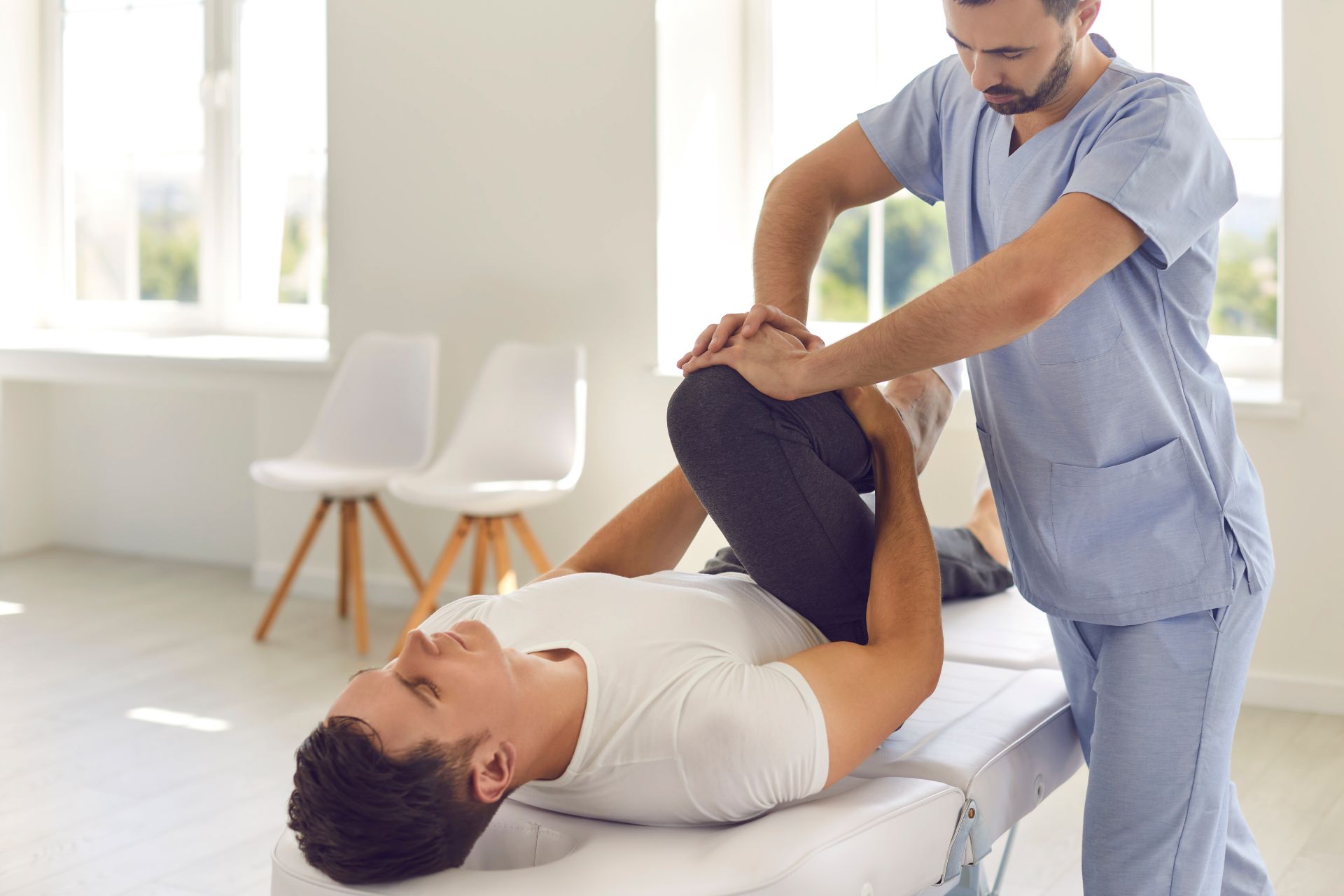
In the first week following knee surgery, specific range of motion exercises are crucial for promoting healing and restoring mobility. These typically include gentle heel slides, where the patient lies flat while sliding their heel towards their buttocks to flex the knee gradually; straight leg raises that engage quadriceps strength without putting excessive strain on the joint; and seated knee extensions performed in a chair to encourage full extension of the leg. Additionally, ankle pumps can be incorporated to enhance circulation and prevent blood clots, while wall slides may also be utilized for controlled bending under supervision. It is essential that these exercises are performed within a pain-free range to avoid exacerbating any swelling or discomfort post-surgery. Each movement should focus on improving flexibility, stability, and overall functional recovery as part of a comprehensive rehabilitation protocol initiated by healthcare professionals.

Weight-bearing status during rehabilitation varies significantly among different types of hip replacement surgeries, such as total hip arthroplasty (THA) and partial hip replacement. In THA, where both the acetabulum and femoral head are replaced, patients may initially follow a protocol that allows for limited weight-bearing on the operated limb to facilitate proper healing of soft tissues and ensure stability of the implant. Conversely, in cases involving hemiarthroplasty or resurfacing procedures, where only part of the joint is replaced, some protocols might permit more immediate weight-bearing activities due to less extensive surgical trauma. The prescribed weight-bearing status often hinges on factors like bone quality, individual comorbidities such as osteoporosis or obesity, patient age demographics, surgeon preference based on clinical guidelines like those from orthopedic associations, and overall functional goals post-surgery. As rehabilitation progresses through phases—initial mobilization followed by gradual increases in load bearing—the emphasis remains on optimizing recovery while minimizing risks associated with mechanical failure or dislocation of prosthetic components amidst varying degrees of activity levels tailored to each specific type of procedure performed.

The transition from passive to active stretching post-shoulder surgery is influenced by several critical criteria, primarily focusing on the patient's range of motion (ROM), pain levels, inflammatory response, and overall functional mobility. Clinicians evaluate whether the patient has achieved adequate joint stability and strength in surrounding musculature before initiating active engagement. Key indicators include a reduction in postoperative swelling and tenderness during rehabilitation exercises, as well as observable improvements in shoulder flexion, abduction, internal rotation, and external rotation. Additionally, assessments of proprioception—particularly neuromuscular control—and coordination play vital roles in ensuring that the patient can safely perform self-directed movements without compromising surgical repairs or exacerbating muscle imbalances. Ultimately, adherence to evidence-based protocols regarding timelines for transitioning to active stretching ensures optimal healing while minimizing risks associated with complications such as adhesive capsulitis or rotator cuff re-tear.

Immediately following ankle reconstructive surgery, effective modalities for managing pain and swelling include cryotherapy, compression therapy, elevation of the limb, and pharmacological interventions. Cryotherapy utilizes cold packs or ice baths to induce vasoconstriction, subsequently reducing edema and alleviating nociceptive pain through decreased inflammatory mediators. Compression garments or wraps enhance venous return while mitigating lymphatic overload, thus minimizing postoperative swelling. Elevating the affected ankle above heart level promotes optimal fluid drainage from periarticular tissues by utilizing gravity’s effects on hydrostatic pressure within interstitial spaces. Additionally, analgesics such as nonsteroidal anti-inflammatory drugs (NSAIDs) are crucial in controlling acute post-surgical discomfort and curtailing inflammation progression at surgical sites. These combined approaches work synergistically to foster a favorable healing environment during the critical initial recovery phase after reconstructive procedures involving ligaments or tendons around the ankle joint complex.

Gait training sessions for patients recovering from total joint arthroplasty should typically be scheduled two to three times per week during the first month post-surgery. This frequency allows for optimal rehabilitation, facilitating improvements in ambulation, proprioception, and overall mobility while addressing any gait abnormalities that may arise due to surgical intervention. During these sessions, physical therapists can focus on enhancing weight-bearing capabilities and limb coordination through targeted exercises aimed at restoring balance and strength in the affected extremity. Additionally, incorporating assistive devices like walkers or crutches may further aid in maintaining safety throughout recovery as patients gradually progress toward independent walking without support. Consistent engagement in these therapeutic interventions is crucial for maximizing functional outcomes and minimizing complications such as stiffness or muscle atrophy following surgery.

Understanding Physical Therapy Protocols in Post-Surgical Recovery

Understanding Physical Therapy Protocols in Post-Surgical Recovery

Contact Us

Alpine Physical Therapy and Fitness

  • Address: 3130 Alpine Rd Suite 370, Portola Valley, CA 94028
  • Phone: (650) 242-0777
  • Email: alpineptfit@mail.com

© Copyright - All Rights Reserved